This section contains real surgical and clinical images shared by Dr. Sejal Kulkarni for medical transparency and educational purposes.
Some images depict intra-operative procedures and surgical specimens. They are shown to demonstrate clinical skill and real patient outcomes — not for general viewing.
By proceeding, you confirm that you are viewing this content voluntarily and understand its clinical/educational nature.
Real outcomes. Real expertise. These case studies reflect Dr. Sejal Kulkarni's commitment to minimally invasive, patient-first surgical care.
Contains photographs of the excised fibroid specimens and post-op scar results. Gated for comfort.
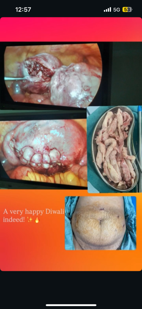
Large intramural fibroid separated from healthy myometrium using precision energy devices.
Multi-layer closure with high-tensile, knotless barbed sutures restores muscular strength.
Tissue extracted safely through a 10mm keyhole port — no open abdominal incision.
Three tiny keyhole port marks vs. a traditional 10–15cm open-surgery scar.
Discharged within 2 days, complication-free, full symptom resolution.
Fibroid Separation: The intramural fibroid was carefully enucleated. Precise haemostasis was maintained throughout using bipolar coagulation, preventing blood loss and preserving uterine tissue thickness.
Meticulous Suturing: Standard three-layer closure was performed to guarantee strong scar integrity, minimizing risks of uterine rupture in subsequent pregnancies. Morcellation was carried out within an extraction bag for safety.
Laparoscopic Myomectomy (fibroid removal)
32-year-old patient with a large intramural uterine fibroid causing heavy menstrual bleeding and pelvic pain
Minimally invasive laparoscopic technique — 3 small ports (<1cm incisions). Fibroid located, excised, and uterus meticulously sutured laparoscopically for complete fertility preservation.
Fertility-preserving approach. No open surgery. No large scar. Uterus remains intact and healthy.
Displays photograph of the removed uterus. Gated for comfort.
Removed uterus specimen showing benign fibroids and changes from severe prolapse — confirms clinical need for excision.
Entire procedure through the vaginal canal — no cut or stitch on the abdomen. Completely scarless recovery.
Pelvic floor muscles and ligaments reconstructed to restore positioning and prevent future prolapse.
Vaginal Hysterectomy — Natural Orifice Surgery (no external incision)
60-year-old post-menopausal female with uterine prolapse — uterus had descended causing significant discomfort and functional difficulty
Vaginal route — no abdominal incision whatsoever. Entire procedure performed through the natural vaginal orifice; pelvic floor reconstructed for long-term support.
The gold standard for uterine prolapse in elderly patients — minimal blood loss, no visible scar, faster healing, immediate relief.
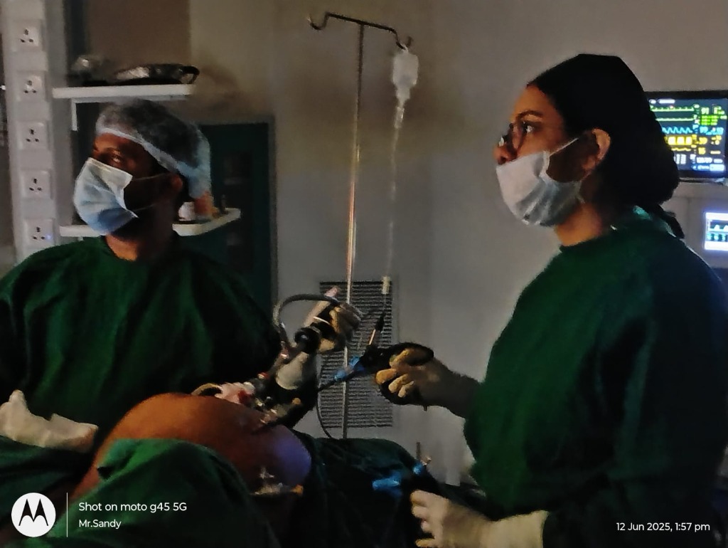
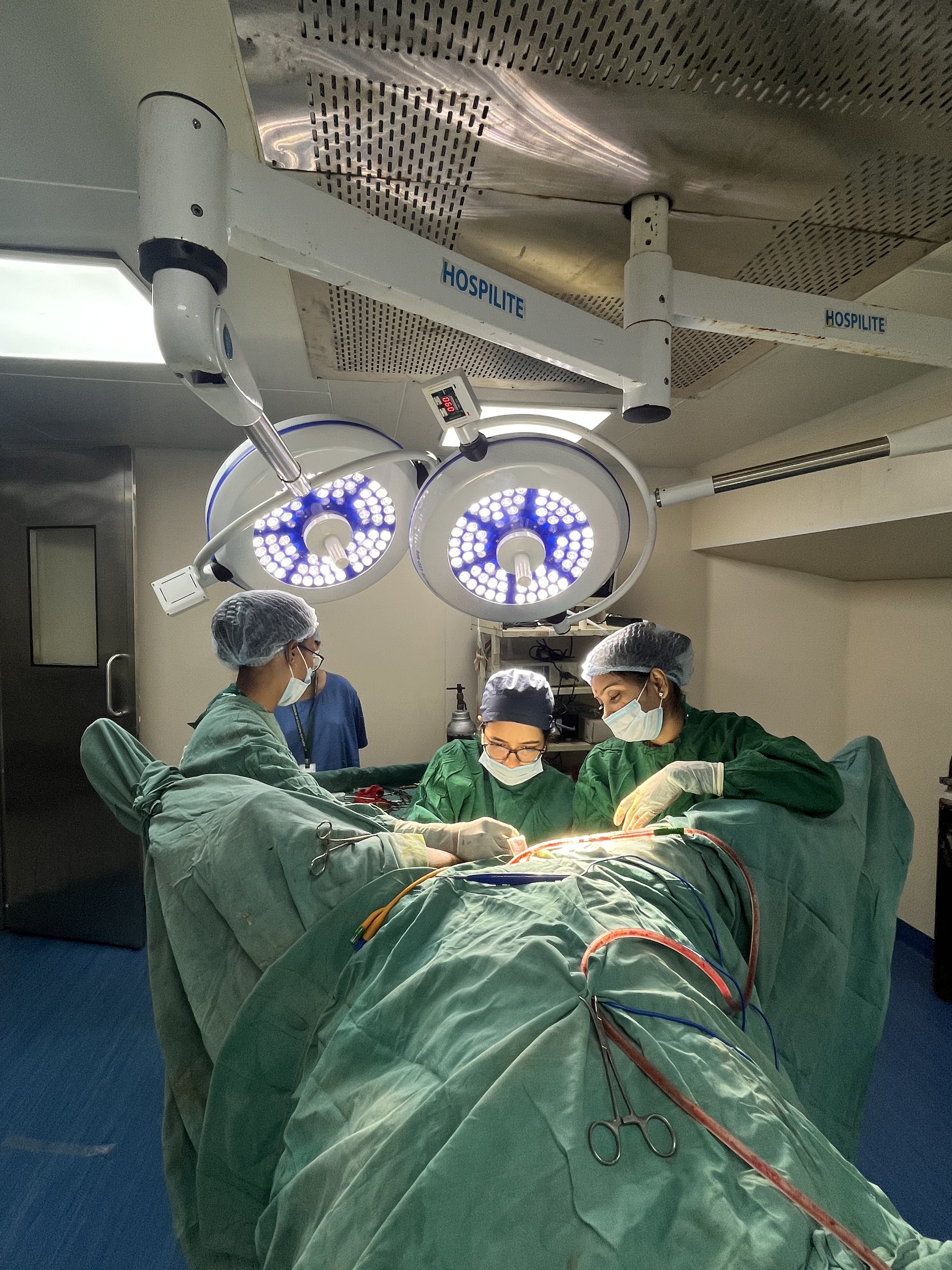
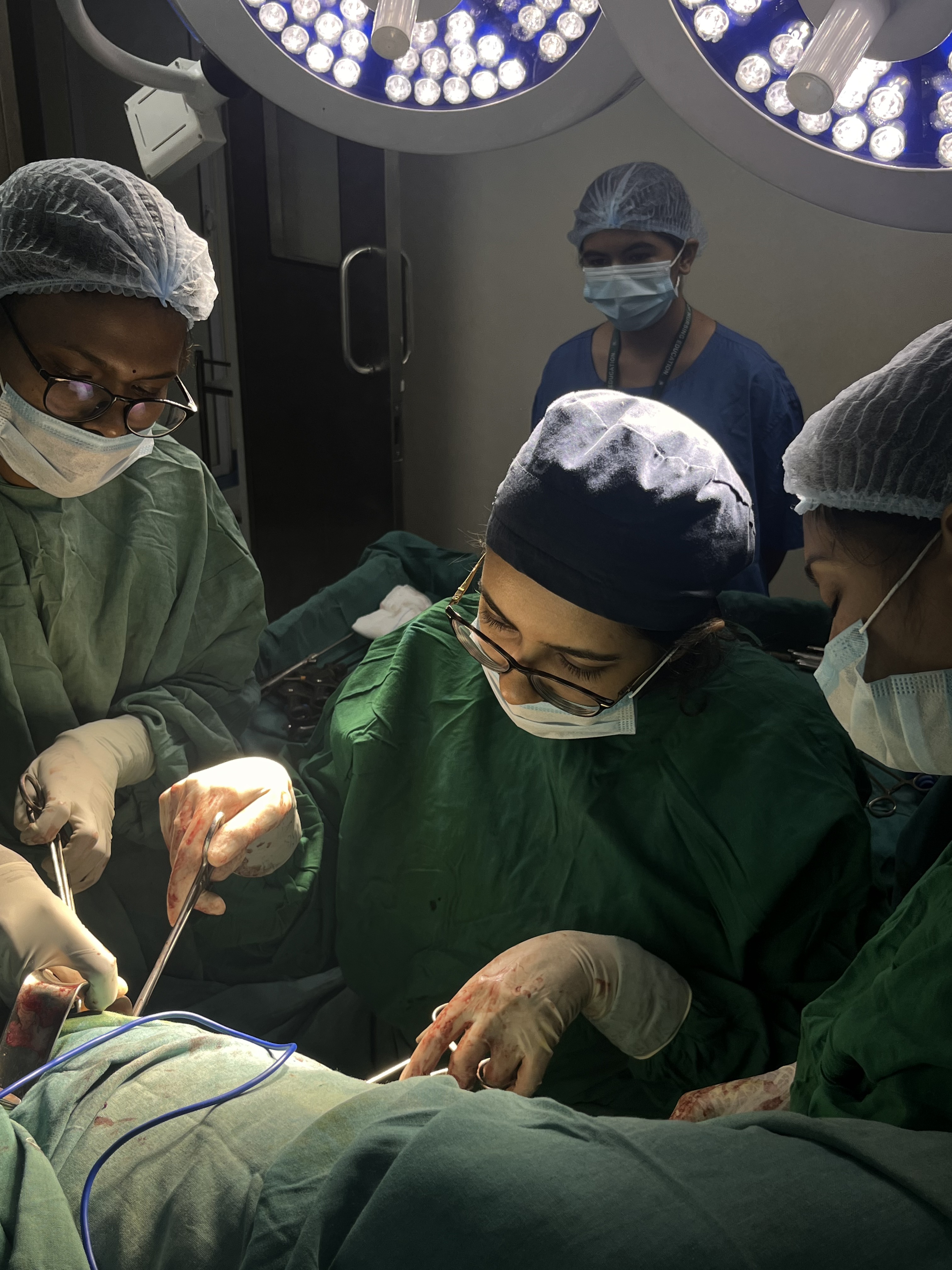
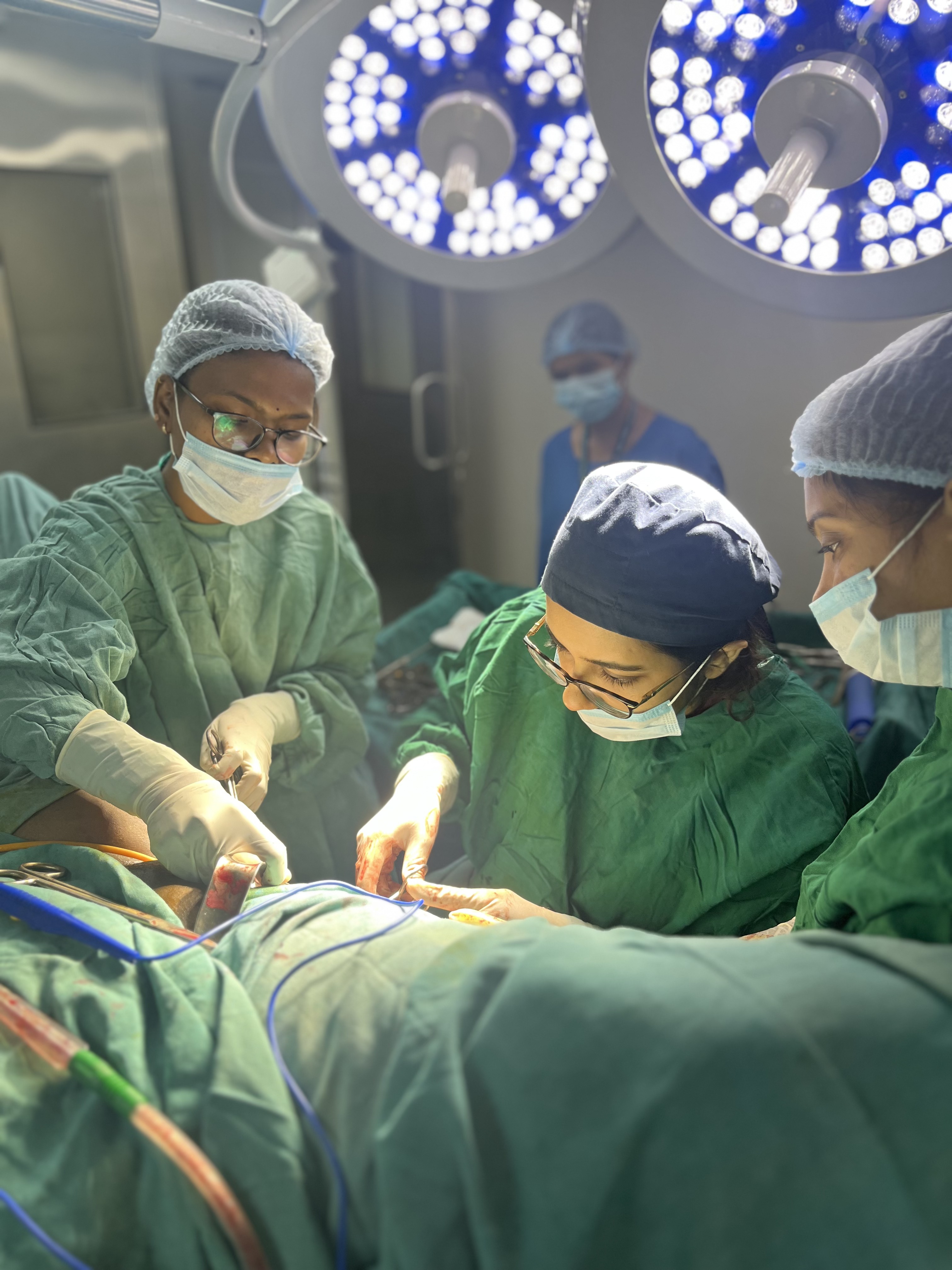
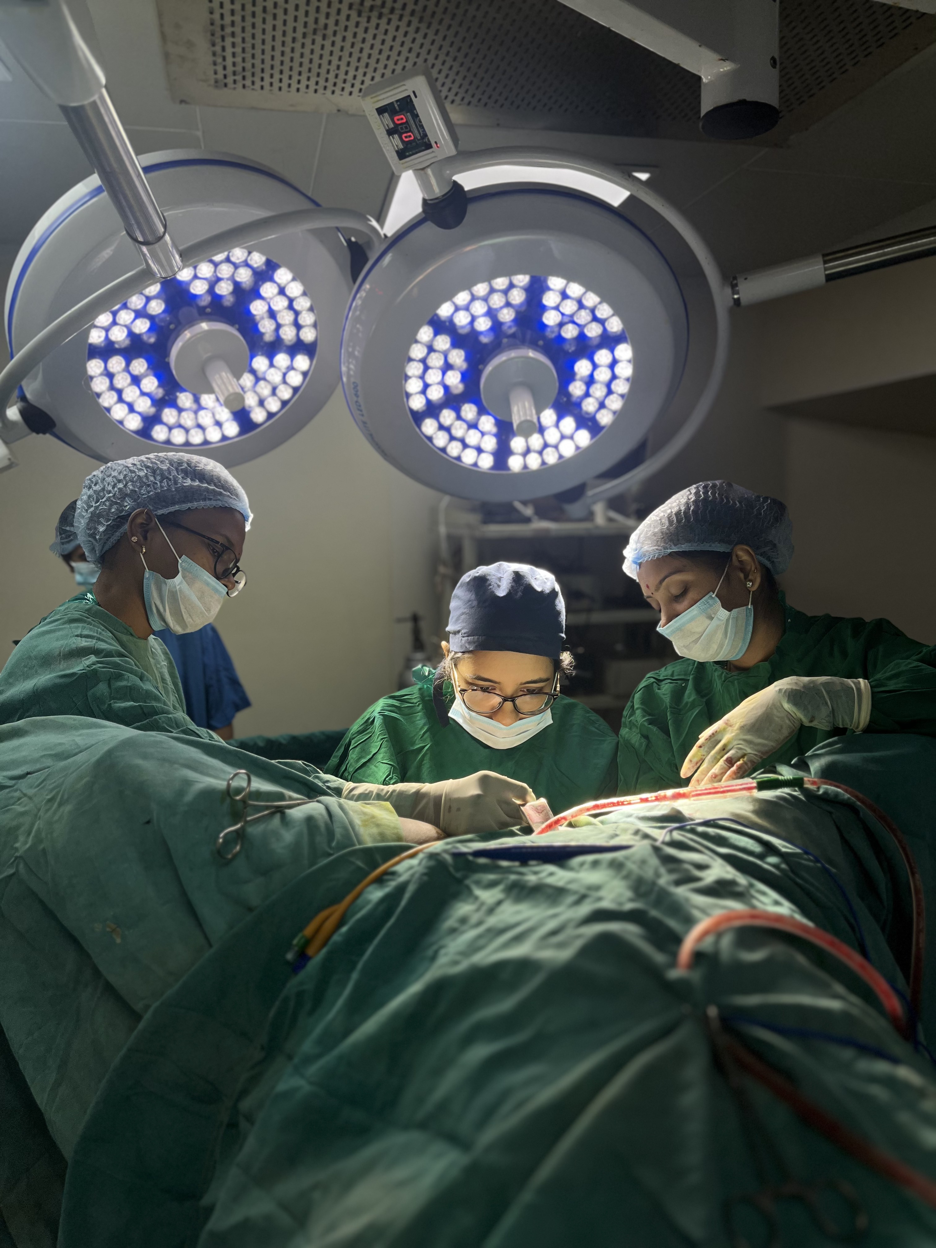
Fully equipped modular operating theater with advanced high-definition endoscopy/laparoscopy stacks.
Continuous intra-operative monitoring (ECG, SpO2, NIBP, EtCO2) and anesthesia delivery systems for maximum patient safety during long laparoscopic procedures.
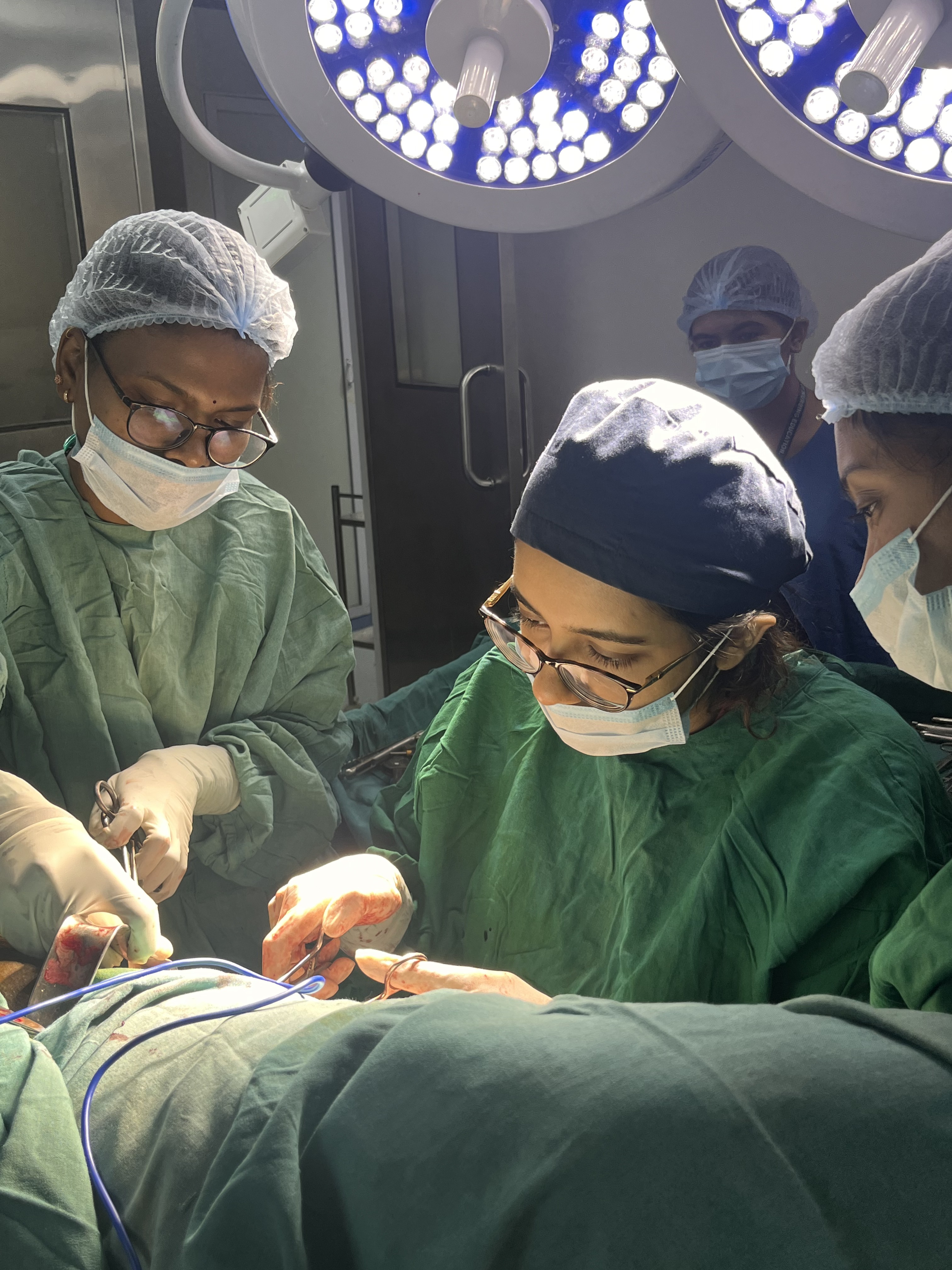
Led by Dr. Sejal Kulkarni with senior consultant anesthesiologists, scrubbing assistants, and circulating nurses specializing in laparoscopic pathways.
Every surgical case is unique. Dr. Sejal will assess your condition in detail and advise the safest, most minimally invasive approach for you.